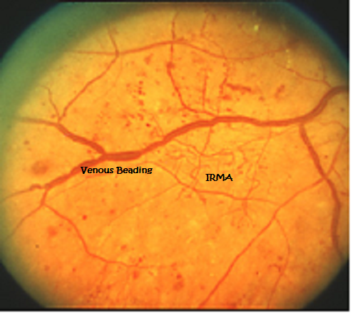
Classification of NPDR is based on clinical findings manifested by visible features, including microaneurysms, retinal hemorrhages, intraretinal microvascular abnormalities (IRMA), and venous caliber changes ( Figure 1), while PDR is characterized by the hallmark feature of pathologic preretinal neovascularization ( 3). DR falls into 2 broad categories: the earlier stage of nonproliferative diabetic retinopathy (NPDR) and the advanced stage of PDR. However, diabetes-induced changes also occur in nonvascular cell types that play an important role in the development and progression of DR, albeit in unison with the vasculature. DR currently affects almost 100 million people worldwide and is set to become an ever-increasing health burden, with estimates between 19 showing that DR-related visual impairment and blindness increased by 64% and 27%, respectively ( 6).īased on their obvious manifestations during DR progression, microvascular lesions have been utilized as the major criteria for evaluating and classifying the retina in DR. Although some reports suggest that the incidence of visual impairment from DR has decreased in recent years in the US largely due to improvements in systemic control ( 5), DR is a burgeoning problem globally. Diabetic retinopathy (DR) is the most common microvascular complication of diabetes ( 4). Patients with diabetes suffer many life-limiting and life-threatening complications, including macrovascular-related stroke, ischemic heart disease, and peripheral artery disease and/or microvascular-related retinopathy, neuropathy, and nephropathy. Type 2 diabetes (T2D) in particular has already attained epidemic levels, while type 1 diabetes (T1D) is increasing in incidence ( 3). The global prevalence of diabetes mellitus is predicted to increase dramatically in the coming decades, from an estimated 382 million in 2013 to 592 million by 2035 ( 1, 2). The clinical challenge of diabetic retinopathy These advances, together with approaches embracing the potential of preventative or regenerative medicine, could provide the means to better manage DR, including treatment at earlier stages and more precise tailoring of treatments based on individual patient variations. This knowledge is leading to identification of new targets and therapeutic strategies for preventing or reversing retinal neuronal dysfunction, vascular leakage, ischemia, and pathologic angiogenesis.

Recent work indicates that diabetes markedly impacts the retinal neurovascular unit and its interdependent vascular, neuronal, glial, and immune cells. However, additional therapeutic options are needed that take into account pathology associated with vascular, glial, and neuronal components of the diabetic retina. Treatments for the vision-threatening complications of diabetic macular edema (DME) and proliferative diabetic retinopathy (PDR) have greatly improved over the past decade. Diabetic retinopathy (DR) causes significant visual loss on a global scale.


 0 kommentar(er)
